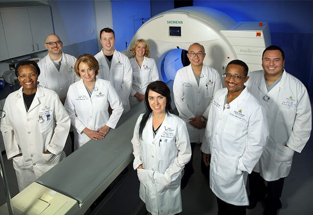
Positron emission tomography (PET) is a specialized radiology procedure that uses molecular imaging to track and trace both normal and abnormal conditions. PET is the most advanced nuclear imaging technique, allowing for high-resolution imaging and absolute quantification of biologic mechanisms. PET imaging is also used to follow the progress of patients undergoing treatment. While PET is most commonly used in the fields of neurology, oncology and cardiology, applications in other fields are currently being studied.
Patient Care
Our PET center team performs more than 4,300 procedures each year. Our subspecialty-trained radiologists manage and interpret every PET scan. These advanced, high-resolution images allow our team and clinical partners access to both the structure and function of organ systems within the body. Our state-of-the-art equipment and technology serve as the backdrop to our unwavering commitment to excellent patient care and attention to detail.
PET scans are often used to help diagnose cancer, detect the spread of cancer to other parts of the body, or measure the effectiveness of cancer treatment. Other uses include diagnosing diseases of the brain and heart, such as Alzheimer's disease, Parkinson's disease, Huntington's disease, epilepsy, stroke, inflammatory heart disease or coronary artery disease.
The Next Generation: PET/CT
Recent technical advances have resulted in the combination PET scanners together with X-ray CT in a single camera system. These hybrid systems yield maximum structural and biological information with a single noninvasive imaging study, combining the power of PET and CT. Our scans help diagnose, treat and evaluate various conditions and diseases
Fast Test Results
Patients can access their test results within three days of their scans through MyChart. MyChart is a secure website that provides the most up-to-date medical information available to you about your Johns Hopkins care and connects you to your health care team. MyChart is available in English and Spanish. To sign up for MyChart, you will receive your unique activation code during your appointment to create your new MyChart user ID and password.
Education
Our division offers a clinical PET/CT fellowship program at Johns Hopkins University School of Medicine. The one-year fellowship program offers specialized training in both clinical and research aspects of positron emission tomography/computed tomography (PET/CT). Established in 1999, this ACGME-accredited fellowship is directly related to the nuclear medicine residency program.
Locations
PET/CT scans are available at The Johns Hopkins Hospital, Johns Hopkins Medical Imaging in Bethesda, and Johns Hopkins Bayview Medical Center. View all location information or download a campus map of The Johns Hopkins Hospital.
PET Center Special Services
Types of scans we perform:
- PET/CT FDG Oncologic Imaging, using the metabolic biomarkers 18F-FDG to help diagnose cancer and detect the spread of cancer to other organs, as well measure the effectiveness of anti-cancer therapy.
- PET/CT Sodium Fluoride Imaging, using the F18-NaF biomarker, a highly sensitive bone-seeking PET tracer, to detect skeletal abnormalities.
- PET/CT FDG Brain Imaging, used to evaluate for cognitive impairment, dementia and seizure disorders.
- PET/CT AMYLOID Imaging, using biomarkers such as F18-Florbetaben and F18- Florbetapir used to estimate amyloid plaque density in patients with cognitive impairment, Alzheimer’s disease and other causes of cognitive decline.
- PET/CT Myocardial Viability Imaging, using 18F-FDG to assess myocardial viability in patients with advanced coronary artery disease and left ventricular dysfunction.
- PET/CT Myocardial Perfusion Imaging, using N-13 ammonia provides high-sensitivity detection of occlusive coronary artery disease, evaluation of the effectiveness of therapeutic interventions, and evaluation of quantitative myocardial flow reserve.
- PET/CT Inflammation and Infection Imaging, using F18-FDG for detection of sarcoidosis, large-vessel vasculitis, implant related infections, and fever of unknown origin.
- PET/CT Ga68 Dotatate Imaging of somatostatin receptors for highly sensitive detection and treatment follow-up of neuroendocrine tumors.